Visualizing electrical activity of the heart
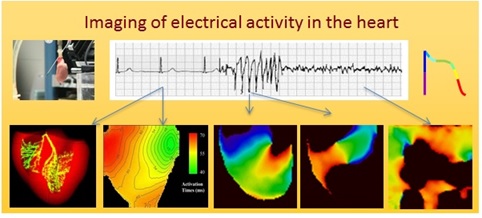
The field of cardiac electrophysiology has focused on gaining a better understanding of the electrophysiological properties of healthy and diseased hearts to gain important insights into the mechanisms of life threatening cardiac arrhythmias, which can cause heart attacks or, in extremely dangerous cases, sudden cardiac death. Optical mapping allows for the visualization of the electrical activities in myocardial tissues with high spatial and temporal resolution. Optical mapping has been widely used in our laboratory to visualize, predict and control the onset of abnormal cardiac rhythms. Over the past years, we further developed technical aspects of the optical mapping system, as well as improved post-process analysis of the acquired data. Specifically, we mastered simultaneous recording of electrical activity of the heart together with intracellular calcium cycling. We also modified the optical mapping system to visualize electrical activity from a very small spatial region. In addition, we incorporated real-time closed loop control that allows us to perform a unique computer-controlled pacing of the heart. All the above mentioned (and other) improvements allow us the use of optical mapping system to tackle a wide spectrum of cardiac electrophysiological applications.
- M. Liu, H. Liu, P. Parthiban, G.-J. Kang, G. Shi, F. Feng, A. Zhou, L. Gu, C. Karnopp, E. G. Tolkacheva, S.C. Dudley Jr. “Inhibition of the unfolded protein response reduces arrhythmic risk after myocardial infarction”, J Clin Invest. 2021 Sep 15;131(18):e147836. doi: 10.1172/JCI147836.
- Molly E. Kupfer, Wei-Han Lin, Vasanth Ravikumar, Ling Gao, Megan Lenz, Didarul Bhuiyan, Jeffrey Ai, DeWayne Townsend, Jianyi Zhang, Elena G. Tolkacheva, and Brenda M. Ogle. In situ expansion, differentiation and electromechanical coupling of human cardiac muscle in a 3D bioprinted, chambered organoid, Circ Res. 2020 Mar 31. doi: 10.1161/CIRCRESAHA.119.316155.
- W. Lee*, K. Kulkarni*, E. G. Tolkacheva, Benchtop optical mapping approaches to study arrhythmias, IEM book chapter, 2018.
- Kulkarni K., Xie X., Fernandez de Velasco E.M., Carlblom N., Wickman K., Tolkacheva E.G., The influences of the M2R-GIRK4-RGS6 dependent parasympathetic pathway on electrophysiological properties of the mouse heart, PLOS ONE, 2018.
- Anderson A., Kulkarni K., Fernandez de Velasco E.M., Carlblom N., Xia Z., Xie X., Nakano A., Martemyanov K., Tolkacheva E.G., Wickman K., Expression and relevance of the G protein-gated K+ channel in the mouse ventricle, Scientific Reports, 2018.
- S. W. Lee*, A. Anderson*, P. A. Guzman, A. Nakano, E. G. Tolkacheva*, K. Wickman*, Atrial GIRK channels mediate the effects of vagus nerve stimulation on heart rate dynamics and arrhythmogenesis, Front Physiol. 2018 Jul 19;9:943. doi: 10.3389/fphys.2018.00943.
Electrical activity in a 3D-printed Chambered Organoid
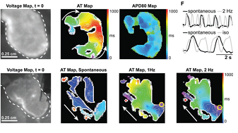
We used optical mapping to record spontaneous electrical activity of a 3D-pronted chambered organoid (left) and electrical activity during pacing.
- Molly E. Kupfer, Wei-Han Lin, Vasanth Ravikumar, Ling Gao, Megan Lenz, Didarul Bhuiyan, Jeffrey Ai, DeWayne Townsend, Jianyi Zhang, Elena G. Tolkacheva, and Brenda M. Ogle. In situ expansion, differentiation and electromechanical coupling of human cardiac muscle in a 3D bioprinted, chambered organoid, Circ Res. 2020 Mar 31. doi: 10.1161/CIRCRESAHA.119.316155.